Educational Content
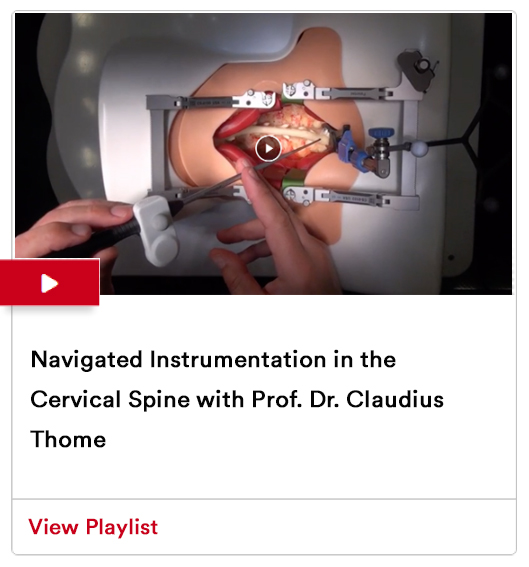
This playlist features 3 videos describing the evolving techniques for complex cervical spine surgeries.
Featured Case Studies
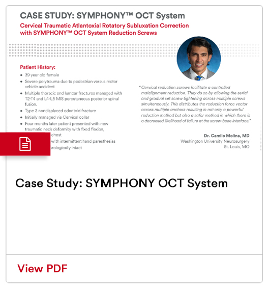
Cervical Traumatic Atlantoaxial Rotary Subluxation Correction with SYMPHONY OCT System Reduction Screws
Dr. Camilo Molina presents a complex cervical trauma case and how the SYMPHONY OCT System Reduction Screws supported the successful patient outcome.
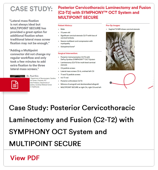
Posterior Cervicothoracic Laminectomy and Fusion (C2-T2) with SYMPHONY OCT System and MULTIPOINT Secure
Dr. Paul Kim presents a 73 year old patient with significant cervical stenosis C2-T1 with loss of cervical lordosis and how surgical intervention using the SYMPHONY OCT System supported positive patient outcomes.
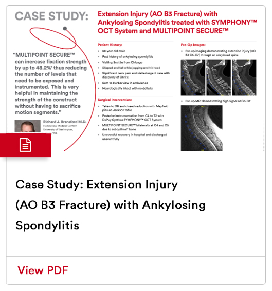
Extension Injury (AO B3 Fracture) with Ankylosing Spondylitis treated with SYMPHONY™ OCT System & MULTIPOINT SECURE™ with Richard Bransford, MD
Dr. Richard Bransford presents a patient with an extension injury and the surgical intervention using SYMPHONY™ OCT System with pre-operative and post-operative imaging.
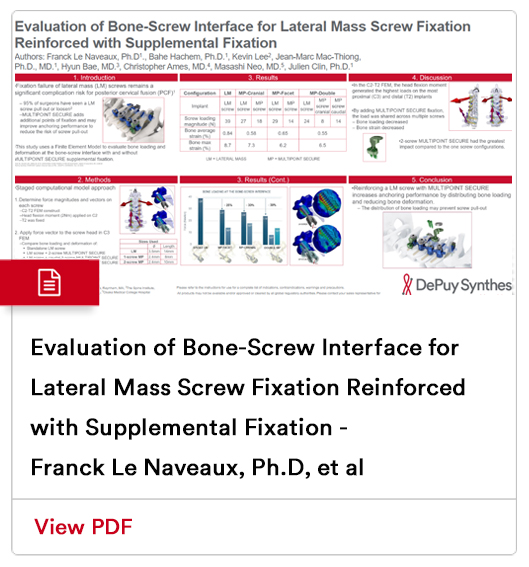
Evaluation of Bone-Screw Interface for Lateral Mass Screw Fixation Reinforced with Supplemental Fixation - Franck Le Naveaux, Ph.D, et al
This case study uses a finite element model to evaluate bone loading and deformation at the bone screw interface with and without MULTIPOINT SECURE supplemental fixation.
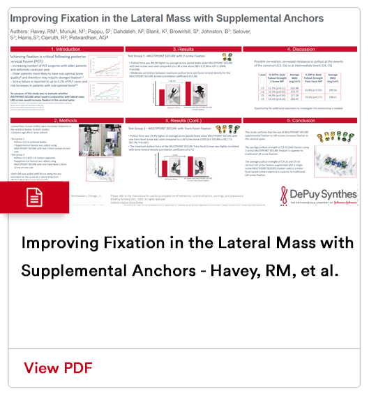
Improving Fixation in the Lateral Mass with Supplemental Anchors - Havey, RM, et al.
This case study confirms that MULITPOINT SECURE used in conjunction with lateral mass screws, increases fixation in the cervical spine.
Additional Cervical Content
Click here to learn more about additional cervical education
offered by Johnson & Johnson MedTech and the Johnson & Johnson Institute.
